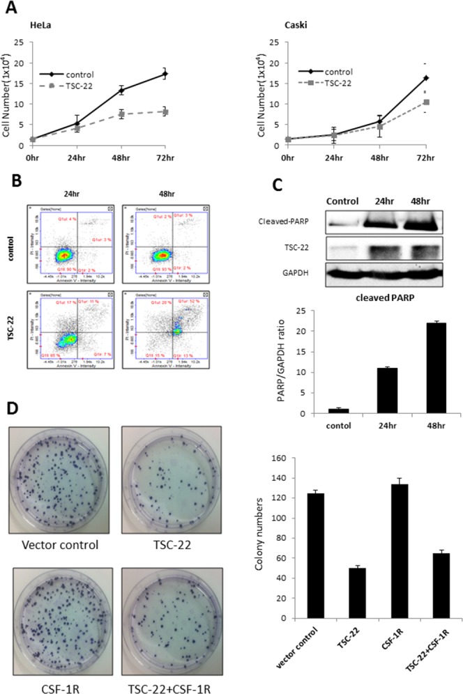
Figure 4. Pro-apoptotic role of TSC-22 in cervical cancer cells.

(A) HeLa and Caski cells were plated in 24-well plates and transiently transfected with pcDNA4 or pcDNA4-TSC-22. The number of surviving cells was counted under a microscope. (B) HeLa cells were transiently transfected with pcDNA4 or pcDNA4-TSC-22. After the indicated time points, AnnexinV/PI double staining was performed to quantify apoptosis of HeLa cells using flow cytometry. (C) Protein level of cleaved PARP was analyzed using western blotting of control and TSC-22 overexpressing cells. (D) TSC-22- or CSF-1R-transfected HeLa cells were re-plated in 100-mm dishes and cultured for 14 days. The number of crystal violet stained colonies was counted.
