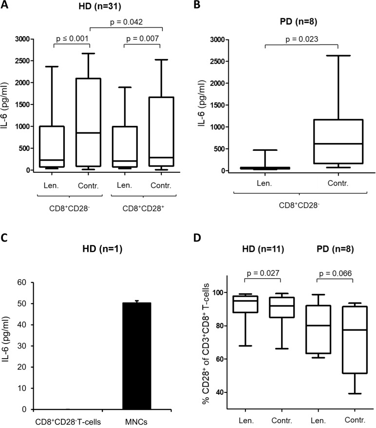
Figure 2. Lenalidomide decreases the IL-6 secretion of MNC.
The supernatants of the incubation-setting MNC with peptide-pulsed DC in the presence of CD8+CD28– T-cells or CD8+CD28+ T-cells and in the presence/absence (Contr.) of lenalidomide were harvested after 12 d and were analyzed by IL-6-ELISA. (A) Shown is the results of 31 HDs. The IL-6 secretion levels in the presence and absence of CD8+CD28– T-cells and lenalidomide (Len., 10 μM) are compared. (B) Shown is the results of 8 patients with PD. The IL-6 secretion levels in the presence of CD8+CD28– T-cells and in presence and absence of lenalidomide (Len., 10 μM) are compared. We used the CD8+CD28– T-cells from patients with PD and the MNC from various HDs. (C) Highly purified CD8+CD28– T-cells and MNC from a HD were incubated for 7 d. Afterwards IL-6 was analyzed in the supernatant by ELISA. (D) The MNC of HDs and patients with PD were incubated with peptide-loaded DC in the presence or absence of lenalidomide (Len., 10 μM) for 12 d. Expanded antigen-specific T-cells were activated with peptide-loaded T2 cells for 48 hrs. For flow cytometry analysis, cells were stained for CD3, CD8, and CD28. The results of CD28–expression on gated CD3+CD8+ T-cells are shown for 11 HDs, and for 8 patients with PD.

