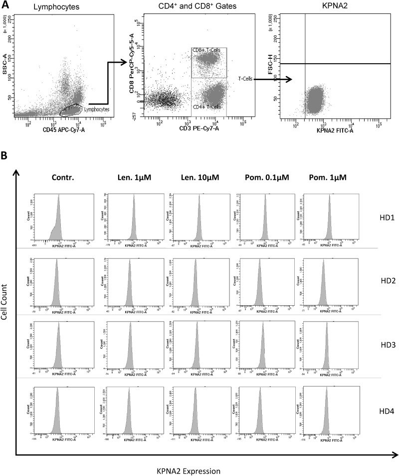
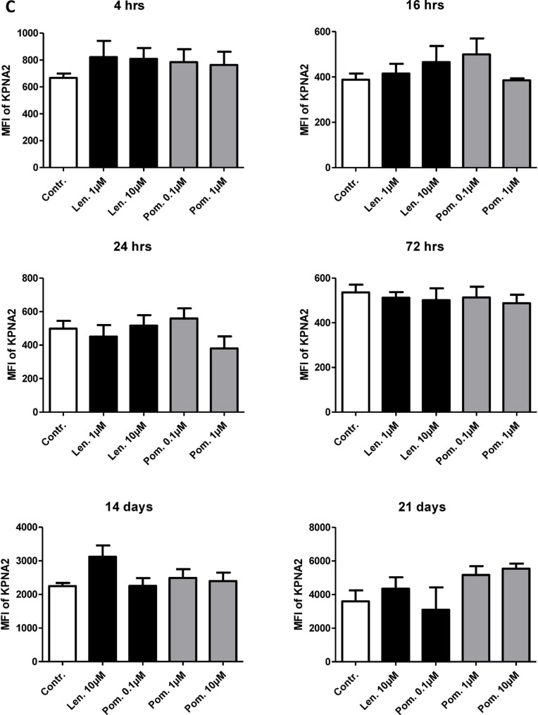
Figure 6. Impact of lenalidomide and pomalidomide on the degradation of the cereblon-binding protein KPNA2.
The MNC of 4 HDs were incubated with peptide-loaded DC in the presence of lenalidomide (1 μM and 10 μM) or pomalidomide (0.1 μM, 1 μM) or without lenalidomide/pomalidomide as the negative control (Contr.). After incubation for 4 hrs, 16 hrs, 24 hrs, 72 hrs, 14 d and 21 d, the cells were stained for CD3, CD8 and KPNA2 and were analyzed by flow cytometry. Figure 6 shows the gating strategy (A) and the KPNA2-expression of CD3+CD8+ T-cells as representative histogram from 4 HDs (after 4 hrs, B) and as cumulative results from 4 HDs (C).


