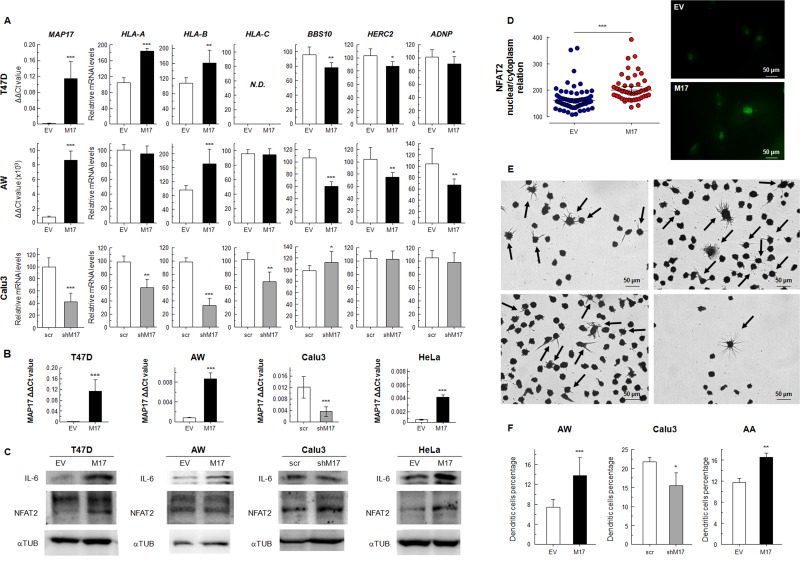
Figure 5.
(A) mRNAexpression levels of HLA-A, HLA-B, HLA-C, BBS10, HERC2 and ADNP in cancer cells transfected for overexpression of MAP17 (T47D, AW) or knockdown of MAP17 with specific shRNA (Calu3). (B) MAP17 expression in human cells transfected to induce its overexpression (T47D, AW, HeLa) or knockdown (Calu3). (C) WB of NFAT2 and IL-6, pro-inflammatory proteins, in cancer cells that overexpress MAP17 (T47D, AW, HeLa) or with its expression downregulated (Calu3). IL-6 is clearly overexpressed in cell lines with higher MAP17 levels, while NFAT2 appears also with higher levels in the cells that overexpress MAP17. (D) NFAT2 nuclear proportion is increased due to MAP17 overexpression. Left) nuclear/cytoplasm relation in AW cells transfected with EV or MAP17 overexpressing vector, according to the fluorescence microscopy images obtained of NFAT2 expression. Right) Fluorescence microscopy of AW EV or AW MAP17 cells. The latter exhibits higher nuclear levels of NFAT2 (E) U937 cells attached to the plate, after being exposed to conditioned cell media (See Methods). Some cells showed the typical form of differentiated dendritic cells (black arrows). (F) Percentage of attached U937 cells for different conditioned media. The percentage of cells with dendritic morphology is higher for cells with higher MAP17 levels. All experiments were repeated a minimum of three independent times in triplicate. All figures include Student's T test for statistical analysis of the data. * = p < 0.05; ** = p < 0.01; *** = p < 0.001.

