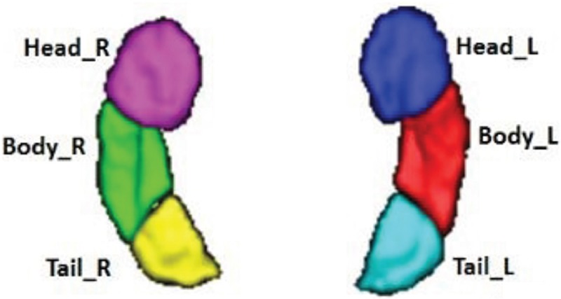
Figure 1. Segmentation of the bilateral hippocampi, which shows the volume rendering of the parcellation results.
Subfields are denoted by different colors. Head_R: right hippocampus head. Head_L: left hippocampus head. Body_R: right hippocampus body. Body_L: left hippocampus body. Tail_R: right hippocampus tail. Tail_L: left hippocampus tail.

