
Figure 2.
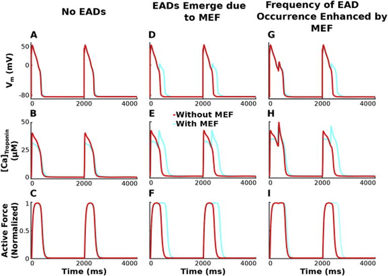
Vm (row 1), [Ca]Troponin (row 2), and active force (row 3) are plotted over time for simulations incorporating HCM-induced ionic remodeling in the absence of myofilament remodeling for a pacing CL of 2000 ms, SL=1.90 μm, and GKr 50% of baseline. Columns 1–3 have progressively enhanced GCaL (3.8, 4.0, 4.2 fold above baseline), illustrating that the degree of repolarization reserve reduction affects the emergence of EADs and the frequency of their occurrence differently for simulations with the bidirectionally coupled model (with MEF; light blue) vs the unidirectionally coupled model (without feedback; red).