
Figure 3.
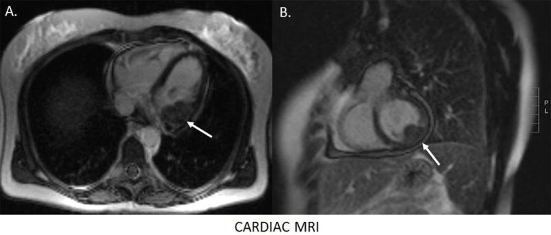
Subsequently, a cardiac MRI with contrast was performed. Multiple cardiac metastatic lesions were identified, the largest in the basal inferolateral left ventricular wall (A, B). The patient’s tumor progressed through treatment with Docetaxel and Gemcitabine with enlargement of the myocardial metastasis and development of a pericardial effusion. The patient developed complete heart block and decided to forego pacemaker implantation, passing away from metastatic lung cancer 6 months after the diagnosis of the cardiac metastases by 18F-FSPG PET/CT. This case highlights the unique advantage of 18F-FSPG PET/CT in tumor imaging. 18F-FSPG is taken up by metabolically active cells via the xC− cystine/glutamate antiporter.1–4 18F-FSPG has a more favorable biodistribution profile than 18F-FDG for detecting malignancy in the liver, bowel, myocardium and brain.5–8 Further investigations in the use of 18F-FSPG in lung cancer and other malignancies are needed to further establish the sensitivity and specificity as well as the role of 18F-FSPG PET/CT in clinical imaging.8–9