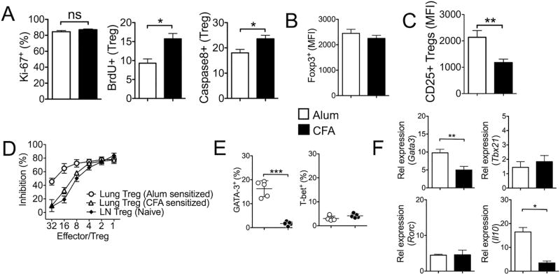
Figure 7. Treg cells from Alum-sensitized mice display more suppressive phenotypes.
(A) Lung infiltrating Foxp3+ Treg cells from Alum- and CFA-sensitized mice were examined for Ki67 expression. Mice were injected with BrdU 24 hours prior to sacrifice. BrdU incorporation of Treg cells was determined by FACS. Active caspase 8 expression was also measured. (B) The level of Foxp3 expression was measured by GFP expression between the groups. (C) Treg cells were stained for surface CD25 expression. (D) Foxp3+ Treg cells were FACS sorted from the lung tissues of Alum- and CFA-sensitized animals. Treg cell suppression assay was performed using CFSE labeled naïve CD4 T cells as described in the Methods. Foxp3+ Treg cells isolated from lymph nodes of naïve animals were used as controls. % inhibition was calculated based on the CFSE dilution of responder CD4 T cells without Treg cells in the culture. (E) Gata3 and T-bet expression was determined by intracellular FACS analysis. (F) Treg cells were FACS sorted and the expression of the indicated genes was determined by real time PCR analysis. The data shown represent the mean ± s.d. of more than two independent experiments. *, p<0.05; **, p<0.01; ns, not-significant.

