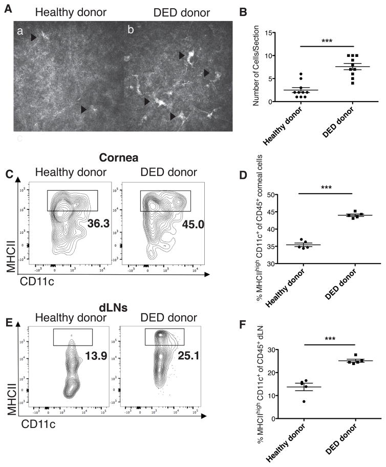
Figure 2. Dry eye disease in donor promotes dendritic cell maturation in graft recipient.
(A) Representative in vivo confocal microscopy (IVCM) images displaying dendritic cells in the cornea of mice with DED and in healthy donors (n=10/group). The size of image is 400 × 400 μm2. (B) Number of dendritic cells, identified as bright dendritiform cells, per section in DED and healthy donor corneas (***p <0.001). (C) Representative flow cytometry plots showing mature CD11c+ MHCIIhigh dendritic cells (DCs) in cornea 14 days post-transplantation. (D) Mean frequencies of mature CD11c+ MHCIIhigh DCs among CD45+ cells in the cornea were assessed using flow cytometry (n=5, ***p<0.001). (E) Representative flow cytometry plots showing mature CD11c+ MHCIIhigh DCs in the draining lymph nodes (dLNs). (F) Mean frequencies of mature CD11c+ MHCIIhigh DCs among CD45+ cells in the dLNs were assessed using flow cytometry (n=5, *p<0.05). p values were calculated using the Student’s t-test and error bars represent standard error of mean.

