
Figure 1.
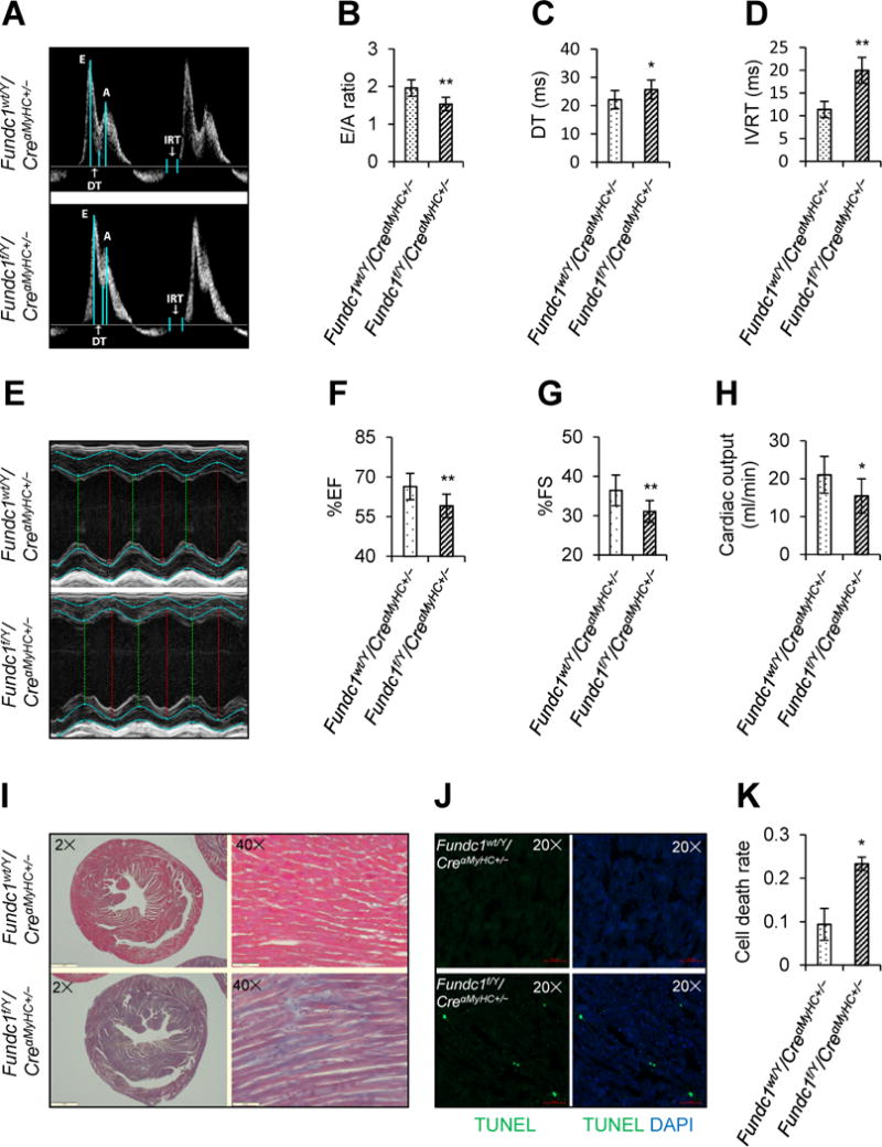
Effects of cardiac Fundc1 deletion on cardiac structure and function. A-H, Echocardiographic assessment was performed in 10-week-old male Fundc1wt/Y/CreαMyHC+/− and Fundc1f/Y/CreαMyHC+/− mice. A, Representative Doppler flow measurements of mitral inflow. B, Ratios of the early (E) to late (A) ventricular filling velocities. C, Deceleration times (DT). D, Isovolumic relaxation times (IVRT). E, Representative images of M-mode echocardiography. F, Ejection fraction (EF). G, Fractional shortening (FS). H, Cardiac output. (Mean ± SD, n = 8 mice per group; *P < 0.05, **P < 0.01 versus Fundc1wt/Y/CreαMyHC+/−.) I, Cardiac fibrosis was revealed by Masson’s trichrome staining of heart paraffin sections. Representative staining is shown (n = 8 mice per group). Scale bars, 1 mm (2×) and 500 μm (40×). J, Representative terminal-deoxynucleoitidyl transferase mediated nick end labeling (TUNEL) staining of heart frozen sections revealed cardiomyocyte apoptosis. Scale bars, 50 μm. K, Statistical analysis of TUNEL staining in J (mean ± SD, n = 8 mice per group; *P < 0.05 versus Fundc1wt/Y/CreαMyHC+/−).