
Figure 4.
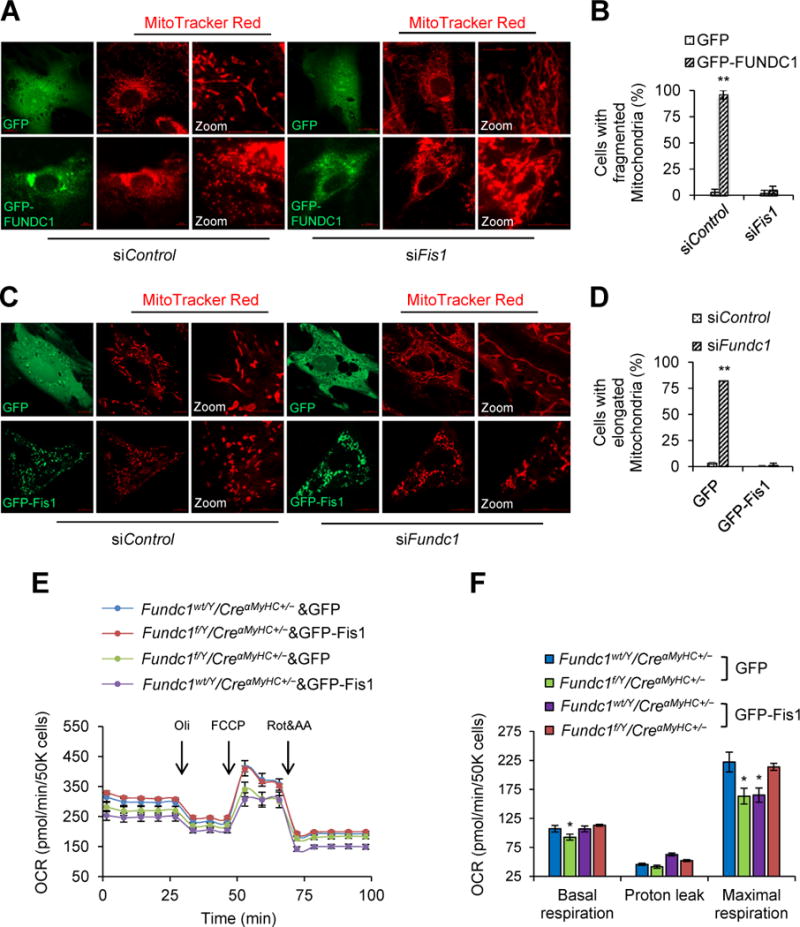
Mitochondrial fission 1 protein (Fis1) is essential for the induction of mitochondrial fission by FUNDC1. A, Representative confocal images of mitochondria in H9c2 myoblasts that were co-transfected with GFP-FUNDC1 plasmid and Fis1 siRNA (siFis1) for 48 h. A scramble siRNA (siControl) was used as gene silence control. Green fluorescent protein (GFP) vector plasmid was used as gene overexpression control. Mitochondria were labeled with MitoTracker Red. Scale bars, 10 μm. B, Quantification of the cells with fragmented mitochondria (mean ± SD, n = 5 independent experiments; 20 cells were quantified per group; **P < 0.01 versus GFP). C, Representative confocal images to show the mitochondrial network in Fundc1-silenced (siFundc1) H9c2 myoblasts with GFP or GFP-Fis1 overexpression. Mitochondria were labeled with MitoTracker Red. Scale bars, 10 μm. D, Quantification of the cells with elongated mitochondria (mean ± SD, n = 5 independent experiments; 20 cells were quantified per group; **P < 0.01 versus siControl). E, Fundc1wt/Y/CreαMyHC+/− and Fundc1f/Y/CreαMyHC+/− neonatal cardiomyocytes were transfected with GFP-Fis1 or control GFP plasmid. Oxygen consumption rate (OCR) was determined using a Seahorse flux analyzer. F, Basal respiration, proton leak and maximal respiration were determined (mean ± SD, n = 5 independent experiments; *P < 0.05 versus Fundc1wt/Y/CreαMyHC+/− & GFP).