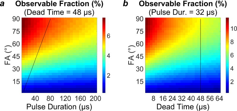
Figure 3.
Plots of the observable fraction of the total longitudinal magnetization. (a) Observable fraction as a function of flip angle (FA) and pulse duration (tp), at a constant TE of 48 µs. (b) Observable fraction as a function of flip angle and TE, at constant pulse duration of 32 µs. Black lines superimposed on the plots illustrate the capabilities of the RF hardware used in the 3T imaging portion of this work.

