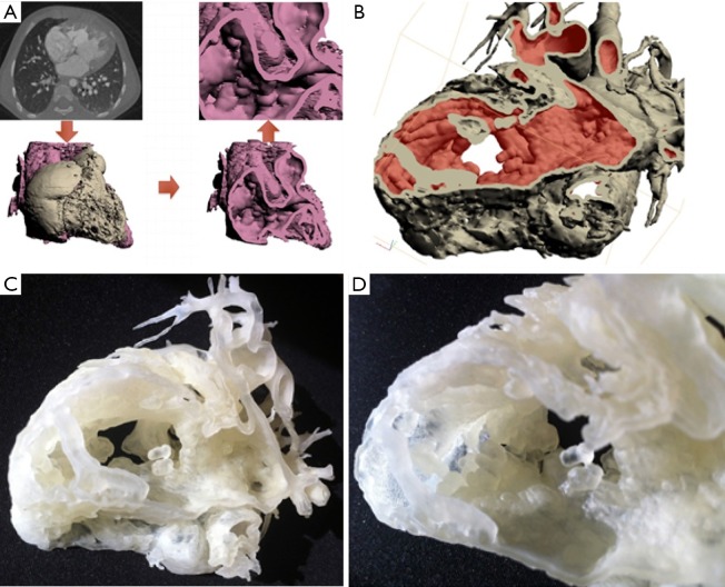
Figure 16.
Paediatric patient with a large ventricular septal defect (VSD) referred for repair surgery. From conventional imaging it was unclear whether to proceed with closing the defect, so 3D printing from gated cardiac CT was used for visualisation. Repair surgery was planned using the 3D printed model and closure of the defect was performed successfully. (A) and (B) 3D mesh demonstrating the VSD generated from gated cardiac CT. (C) and (D) 3D printed model of the VSD used for surgical planning.

