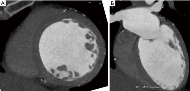
Figure 4.
Dilated cardiomyopathy. (A) Short-axis CT image shows severe dilation of the left ventricle with end-diastolic diameter of 7.1 cm; (B) 4-chamber CT image also shows severe dilation of the left ventricle. The ejection fraction in this patient was 15% and the constellation of findings is consistent with dilated cardiomyopathy. CT, computed tomography.

