Figure 7.

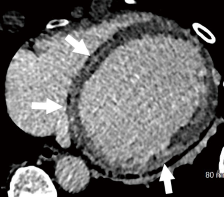
Sarcoidosis. Axial delayed enhancement CT obtained 10 min after contrast in a patient with sarcoidosis shows mid-myocardial delayed iodine enhancement in the septum and the lateral wall (arrows). CT, computed tomography.

Sarcoidosis. Axial delayed enhancement CT obtained 10 min after contrast in a patient with sarcoidosis shows mid-myocardial delayed iodine enhancement in the septum and the lateral wall (arrows). CT, computed tomography.