Figure 9.

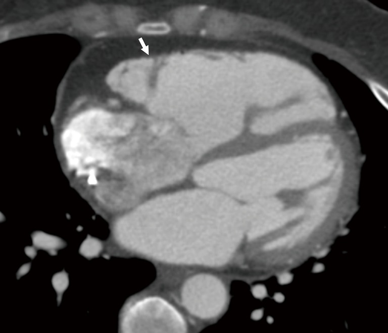
ARVD. Axial CT scan in a patient with ARVD shows dilated right ventricle and presence of fat in the RV free wall (arrow). ARVD, arrhythmogenic right ventricular cardiomyopathy; CT, computed tomography.

ARVD. Axial CT scan in a patient with ARVD shows dilated right ventricle and presence of fat in the RV free wall (arrow). ARVD, arrhythmogenic right ventricular cardiomyopathy; CT, computed tomography.