
Figure 2.
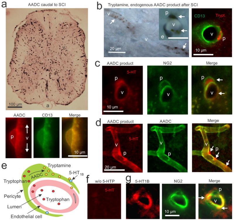
AADC, trace amines and 5-HT1B receptors are co-expressed in pericytes after SCI. (a) Top, immunolabeling with an AADC antibody (black, DAB, upper panel) in a transverse section of a spinal cord caudal to a chronic spinal transection, showing that AADC is widely expressed on capillaries (v), but not arteries (a). Bottom, immunofluorescence for AADC (red) and CD13 (green, pericyte marker) in a lengthwise section of capillary, showing exclusive colocalization of AADC and CD13 in pericytes (p; yellow). (b) Left, DAB immunolabeling of endogenous tryptamine (black) caudal the site of injury, showing dense staining for tryptamine in pericytes (arrows) of capillaries (v), especially in the soma. Inset shows a higher magnification view of a capillary cross-section (scale bar, 10 μm), showing that pericyte (p) cell bodies and processes stain for tryptamine (arrows), but an endothelial cell (e) does not (blue, cresyl-violet stain of the endothelial cell nucleus). Right, immunofluorescent staining for tryptamine (red) and CD13 (green) further showing tryptamine staining in a pericyte. (c) Immunolabeling for the AADC product 5-HT and for the pericyte marker NG2 caudal to a chronic transection injury after pre-treatment with 5-HTP (30 mg/kg, i.p., 25 min prior to fixation). Arrows, staining of 5-HT in NG2-labeled pericytes. (d) This AADC product 5-HT (red) is shown densely accumulated in pericyte cell bodies and processes labelled for AADC (green; at arrows). (e) Schematic of pericyte action on capillaries after SCI, showing diffusion of tryptophan (red) from blood into pericytes, synthesis of tryptamine (yellow) from AADC, and the action of tryptamine on nearby 5HT1B receptors (blue) to constrict the capillary. (f) Immunolabeling for 5-HT as in panels c and d, but without 5-HTP pre-treatment. (g) Immunolabeling for the 5-HT1B receptor and NG2 caudal to the site of injury. Arrows show localization of 5-HT1B receptor on NG2-labelled pericytes, with dense areas of receptor staining on pericyte processes. n = 5 rats tested per condition.