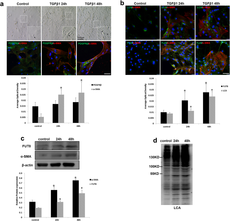
Figure 4.
Core fucosylation was increased during the TGFβ1-induced pericyte-myofibroblast transition in vitro. Primary cultures of pericytes were incubated with TGFβ1 (10 ng/ml) for 24 or 48 h. (a) Representative bright-field images of morphological alterations in pericytes, where black arrows indicate pericytes, and representative images of PDGFRβ (green) and α-SMA (red) staining are shown. (b) Representative images of dual staining for FUT8 (green) and α-SMA (red) and dual staining for LCA (green) and α-SMA (red) staining. (c) PDGFRβ, α-SMA, and FUT8 levels were assessed using Western blot analyses. (d) Lectin blot analyses. Quantification is shown in the lower panel. Scale bar, 50 μm. *P < 0.01, † P < 0.01. * and †indicate the comparison of the control group with the TGFβ1 group; # indicates the comparison of the FUT8 knockdown group with the TGFβ1 group.

