Figure 1.
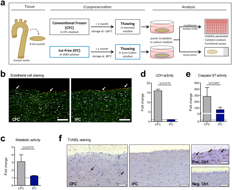
Histological and metabolic characteristics of human aortic tissue after Conventional Frozen Cryopreservation (CFC) or Ice-Free Cryopreservation (IFC). (a) The tissue preparation and analysis is shown schematically. Punches of fresh aortic tissue were made and frozen according to either the CFC or IFC protocols (see methods). Tissue punches were stored at least 1 month in the vapor phase above liquid nitrogen (about −160 °C) or at −80 °C before use, respectively. Tissue samples were thawed and washed according to the CFC or IFC protocols for further analysis. Punches were incubated in cell culture medium and tissue viability (metabolism, apoptosis, and necrosis) and cytokine release were analyzed, and tissue specimens were histologically examined. (b) Human CD31+ staining (yellow) indicates the endothelial cell layer (white arrows) on the intimal side of both CFC and IFC aortic tissue. Nuclei were stained with DAPI (pseudo-colored white); while the extracellular matrix shows green autofluorescence. Scale bars represent 100 µm. (c) Metabolic activity of the tissue was assessed with an MTS assay. (d) Necrosis level was determined by the measurement of lactate dehydrogenase (LDH) activity in conditioned medium (CM). (e) Apoptosis of tissue cells was analyzed via measurement of caspase-3 and -7 activities in the CM. All measurements are normalized to the medium control (dotted line). Data are shown as the mean + SEM (n = 5–7) and analyzed with Mann Whitney test *p < 0.05. (f) TUNEL staining revealed some apoptotic cells (brown nuclei) highlighted by black arrows in the CFC tissue. The DNAse-treated positive control (Pos. Ctrl.), and a negative control (Neg. Ctrl.) are shown for comparison. Scale bars represent 75 µm.

