Figure 2.
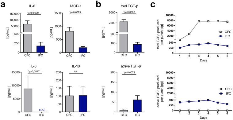
IFC tissue releases lower cytokine and chemokine levels than CFC tissue, however more biologically active TGF-β is present. CFC and IFC aortic tissue punches were incubated in DMEM culture medium for 2 days. The cytokines and chemokines IL-6, MCP-1 IL-8, IL-10 (a) total TGF-β (active and latent form) and active TGF-β (b) were analyzed by ELISA or multiplex bead assay. Data are shown as the mean + SEM (n = 4–8) and analyzed with Mann Whitney test **p < 0.01, ***p < 0.01; n.s.: not significant; n.d.: not detectable. (c) Over a culture period of 6 days, total TGF-β (upper graph) and active TGF-β (lower graph) was measured by ELISA. The absolute amount of TGF-β in pg produced per tissue punch was calculated and a representative kinetic graph is shown.

