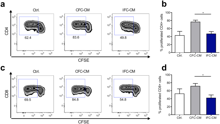
Figure 4.
Soluble factors from CFC tissue but not from IFC tissue amplify αCD3 triggered T cell proliferation. Human CFSE-labeled PBMC were stimulated with a low dose of αCD3 antibody and subjected to CFC or IFC tissue conditioned medium (CM). After 4 days, PBMC were harvested, stained with fluorochrome-labeled human specific antibodies for T cell subset markers and analyzed by flow cytometry. Representative flow cytometry plots show the induction of CD4+ T cell (a) and CD8+ T cell (c) proliferation in PBMC cultures stimulated with either αCD3 alone (Ctrl.; left), or combined with CFC tissue CM (CFC-CM; middle) or IFC tissue CM (IFC-CM; right). Bar graphs show the levels of proliferated CD4+ T cells (b) and CD8+ T cells (d) in summary. Data are shown as the mean + SEM (n = 5–8) and analyzed with one-way ANOVA (Kruskal-Wallis test) *p < 0.05.

