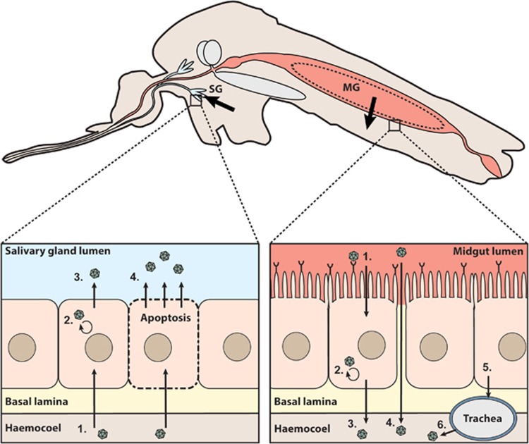
Figure 2.
Schematic overview of the mosquito barriers to arbovirus infection. Schematic longitudinal cross-section of a mosquito. Arrows indicate the passage of virions through the midgut (MG) and salivary gland (SG) barriers. The dashed circle in the midgut represents the peritrophic membrane that is formed after ingestion of blood. Right inset: (i) Infection of midgut epithelial cells via binding to a putative receptor protein. (ii) Virus replication in midgut epithelial cells. (iii) Release of virus via budding from midgut epithelial cells and direct passage through the basal lamina into the haemocoel. (iv) Direct virus passage into the haemocoel through a ‘leaky’ midgut. (v) Virus infection of trachea after budding from midgut epithelial cells. (vi) Budding of virus from the trachea into the haemocoel. Left inset: (i) Infection of the salivary gland epithelial cells after passage through the basal lamina. (ii) Virus replication in the salivary gland cells. (iii) Virus release via budding from salivary gland cells into the salivary gland lumen. (iv) Virus release from the salivary gland cells into the salivary gland lumen via apoptosis.

