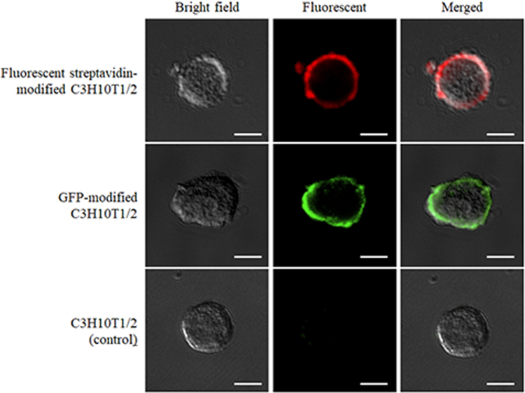
Figure 1.
The typical images of surface-modified C3H10T1/2 cells. Fluorescent streptavidin-modified C3H10T1/2 cells were prepared by adding Alexa Fluor 647 conjugated streptavidin to biotinylated C3H10T1/2 cells. GFP-modified C3H10T1/2 cells were prepared by adding biotin-GFP to avidinated C3H10T1/2 cells. C3H10T1/2 cells (control) were prepared by adding biotin-GFP to unmodified C3H10T1/2 cells after addition of avidin. Scale bars represent 20 μm.

