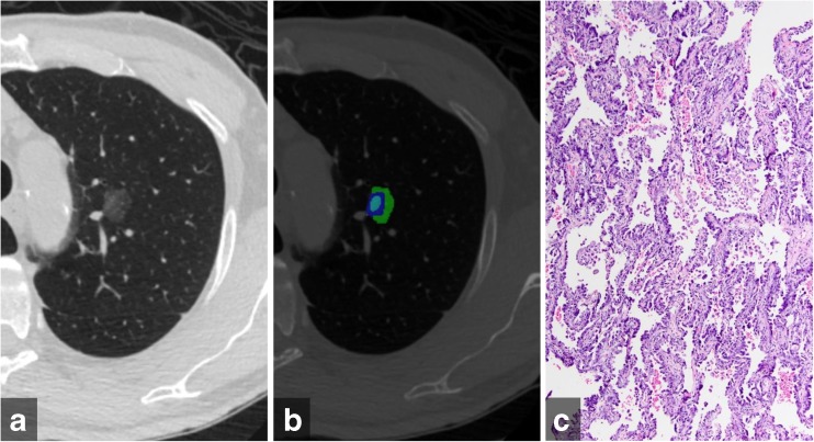
Fig. 1.
(a) Transverse computed tomography image of an adenocarcinoma in situ manifesting as pure ground glass nodule in the left upper lobe (b) Color-coded CANARY output overlay shows components of low risk group (blue-green–cyan) (low risk 99.8%, intermediate risk 0.2%, high risk 0%) (c) histologic image showing purely lepidic tumour growth pattern without areas of invasion (H&E stain, 200x original magnification)

