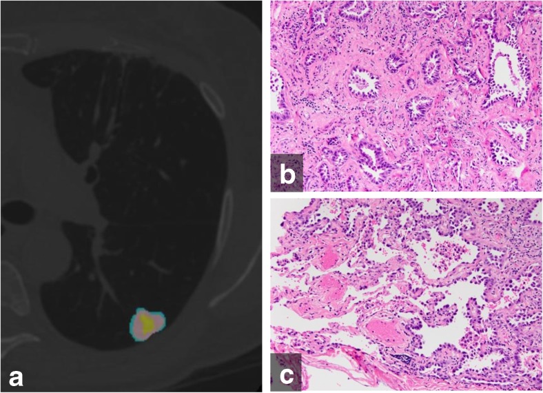
Fig. 4.
(a) Color-coded CANARY output overlay of invasive adenocarcinoma (yellow-pink-cyan) (low risk 32.0%, intermediate risk 66.9%, high risk 1%)(b) histologic image from the central area of the tumour shows the invasive acinar growth pattern (c) histologic image from the tumour periphery showing non-invasive lepidic growth pattern and adjacent uninvolved lung parenchyma (H&E stain, 200x original magnification)

