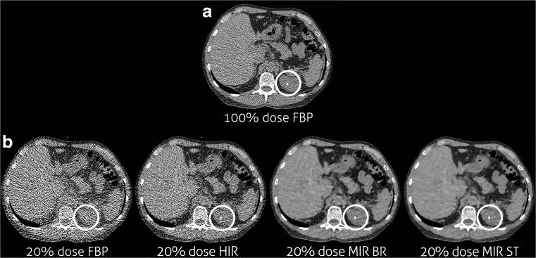
Fig. 1.
Example of decreased sensitivity for stone detection. From left to right the stone at routine dose reconstructed with FBP (a), the FBP reconstruction at the lowest dose level on which the stone was missed (b) and the IR reconstructions (HIR, MIR Body Routine and MIR Soft Tissue) at the same dose level on which the stone is clearly visible

