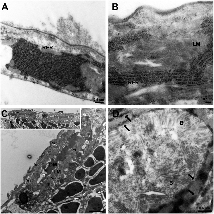
Figure 2.
TEM of CLas and ER dynamics in D. citri infected gut cells. (A) Dense RER in healthy midgut epithelia cell. (B) Rearrangement of rough ER (RER) membranes around the light matter (LM) inside a midgut cell. (C) Liberibacter containing vacuoles (LCVs) showing the LM and the dark matter (DM) inside them. LCVs are located close to midgut basal lamina. Two adjacent LCVs are shown in the inset. LCV borders are indicated with arrows. (D) Zoom-in on one LCV showing mature CLas bacteria (B) in the LM, surrounded by the double membranous ER (arrows).

