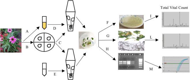
Figure 1.
In vitro model system setting up to study the interaction between Echinacea purpurea plants and their stem/leaves endophytic bacteria. (A) Bacterial endophytes were isolated from the aerial compartment of E. purpurea plants. (B) E. purpurea seeds were provided by the common garden at the “Il Giardino delle Erbe”, Casola Valsenio, Italy and surface-sterilized. (C) Seeds were germinated in De Wit Culture tubes containing 5 ml of Linsmaier & Skoog Medium (LS) including vitamins. After root formation, the seedlings were transferred in Wavin flasks containing 50 ml of LS solid medium, supplemented with 3% sucrose and maintained in a plant growth chamber for a photoperiod of 16 h light a day. (D,E) After about 2 months, five E. purpurea plants were inoculated with 8 × 10⁶ bacterial endophytes isolated from SL compartment of E. purpurea plants (D); five plants were used as control and were inoculated with sterilized saline solution (E). After 45 days, SL and root (R) samples from control and infected plants, were collected separately and sterilized. (F–H) Samples were then, separated in different aliquots (R and SL pooled separately). (I) One aliquot of each tissue was immediately used for the in planta bacterial growth analysis. (L) One aliquot of each tissue was weighed and dried at 60 °C to be used for n-hexane extracts preparation. (M) One aliquot of each tissue was ground to a fine powder in liquid nitrogen and successively used for RNA extraction.

