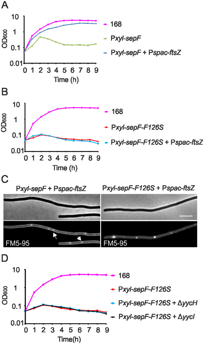
Figure 9.

FtsZ induction suppresses division defect. (A) Growth curves of strain 168 (wild type), strain GYQ215 (amyE::Pxyl-sepF), and strain GYQ77 (amyE::Pxyl-sepF aprE::Pspac-ftsZ) in medium containing 1% xylose to induce SepF and 5 mM IPTG to induce FtsZ expression. (B) Growth curves of strain 168 (wild type), strain GYQ207 (amyE::Pxyl-sepF-F126S ∆sepF) and strain GYQ210 (amyE::Pxyl-sepF-F126S ∆sepF aprE::Pspac-ftsZ) in medium containing 1% xylose to induce SepF-F126S and 5 mM IPTG to induce FtsZ expression. (C) Phase contrast and membrane stain of cells from culture GYQ77 (Fig. 9A), and culture GYQ210 (Fig. 9B). Cells were stained with the membrane dye FM5-95. Scale bar is 5 µm. Normal septa are indicated by arrows. (D) Growth curves of strain 168 (wild type), strain GYQ185 (amyE::Pxyl-sepF-F126S ∆sepF), strain GYQ223 (amyE::Pxyl-sepF-F126S ∆sepF ∆yycH) and strain GYQ224 (amyE::Pxyl-sepF-F126S ∆sepF ∆yycI) in medium containing 1% xylose to induce SepF and SepF-F126S.
