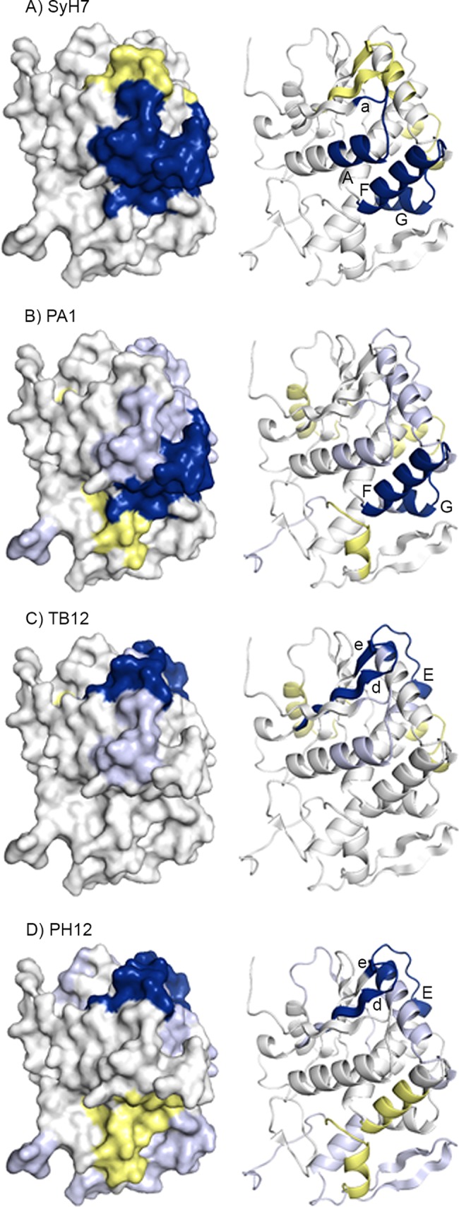
FIG 6.

Visual presentation of the core epitopes of SyH7, PA1, TB12, and PH12 on RiVax. HX protection categories, as defined in the Fig. 5 legend, are shown mapped onto the structure of RiVax for (A) SyH7, (B) PA1, (C) TB12, and (D) PH12. The image is color coded according to the Fig. 5 legend. The locations of β-strands a, d, and e as well as α-helices A, E, F, and G are indicated.
