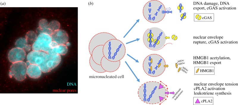
Figure 5.
Candidate inflammatory pathways after micronucleation (illustration by T.J.M.). (a) Immunofluorescence image of a HeLa cell after passage though mitosis in paclitaxel showing multiple micronuclei and nuclear envelopes, in this cased stained for nuclear pores (T.J.M. 2007, unpublished data). (b) Four candidate pro-inflammatory pathways in micronucleated cells. See text for details.

