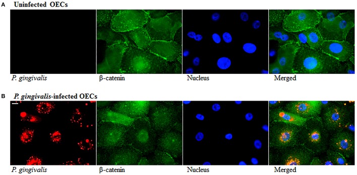
Figure 4.
Altered Distribution of β-Catenin in P. gingivalis-infected cells. OECs were incubated with P. gingivalis at an MOI of 100 for 120 h. (A) Uninfected OECs. (B) P. gingivalis-infected OECs. Uninfected and P. gingivalis-infected cells were fixed, permeabilized, and immunostained with a monoclonal β-catenin antibody (green fluorescence) and for a rabbit P. gingivalis antibody (red-orange fluorescence). Nuclei were stained with DAPI (blue fluorescence). At least three separate fields containing an average of 20 OECs were studied in each of the three independent experiments performed in duplicate. Bar 10 μm.

