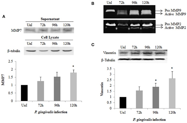
Figure 5.
Activation of MMPs and Vimentin expression in P. gingivalis-infected OECs. OECs were incubated with P. gingivalis at an MOI of 100 for 72, 96, and 120 h. Bradford assay was used to determine the protein concentration of each sample. (A) Supernatants were harvested and precipitated using the TCA method from uninfected and P. gingivalis-infected OECs. The precipitated proteins were immunoblotted and detected using a monoclonal MMP7 antibody. (B) TCA-precipitated proteins were loaded on a SDS-PAGE gel containing 3% of gelatin. The gelatinolytic activity of MMP2 and MMP9 were visualized as clear, non-staining regions of the gel. (C) Cell lysates were extracted from uninfected and P. gingivalis-infected OECs and immunoblotted with a monoclonal Vimentin antibody. The absolute intensities of MMP7 and Vimentin western blot bands were measured using ImageJ software and the relative (normalized) intensities were calculated using β-tubulin as a loading control. Data are shown as the mean ± standard deviation of three independent experiments. *Denotes statistical significance (p < 0.05) according to two-tailed Student's t-test.

