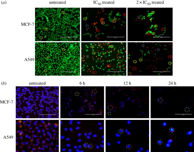
Figure 5.
(a) Fluorescence microscopic images of AO/EB dual staining of untreated, IC50 and 2 × IC50 of Nic-Chi Np's treated MCF-7 and A549 cells. Yellow, blue and red dotted circles represent viable, early apoptotic and late apoptotic cells, respectively. Scale bar, 200 µm. (b) Time-dependent overlay images of untreated and Nic-Chi Np's (IC50) treated MCF-7 and A549 cells stained with Hoechst 33342 (blue) and co-stained with rhodamine B (red). Red doted circle indicates nuclear fragmentation, green shows cytoskeleton compaction. Scale bar, 200, 100 µm.

