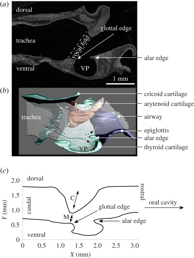
Figure 5.

Sagittal section of a grasshopper mouse larynx. (a) Iodine-stained computer tomographic image. (b) Three-dimensional model of the cartilages and the intralaryngeal airway. (c) Mid-sagittal dimension of the airway of a grasshopper mouse. The white dashed line indicates the dorsal boundary of the airway as it becomes constricted coincident with vocal fold abduction. The glottal opening is close to the ventral surface, with the glottal jet flowing into the supraglottal (epilaryngeal) space to pass over the ventral pouch and impinge upon the alar edge. VP, ventral pouch; M, membraneous portion of the vocal fold; C, cartilaginous portion (supported by vocal process of the arytenoid cartilage) of the vocal fold.
