. 2017 Dec 5;6:Doc15. doi: 10.3205/iprs000117

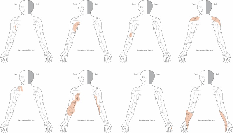
Figure 1. Schematic drawing of dermatomes of the upper limb and hand used for plotting the extent of PNF (illustrated for right arm), exemplified in 8 individuals: areas of tumor affecting the skin (diffuse neurofibroma) or nodular PNF located below the skin are marked in blue. Tumor expansion can continuously infiltrate a skin region without clear reference to one or more dermatomes. Tumors with a smaller area of infiltrated skin are more likely to lie within a dermatome. Some tumors are so large that they alter the contour of the limb. In these areas the color marking of the tumor extension extends beyond the outline of the limb. This simplified schematic drawing does not take into account the highly variable sensory skin innervation of the upper limb as described in detail in anatomical studies [102].

Figure 1