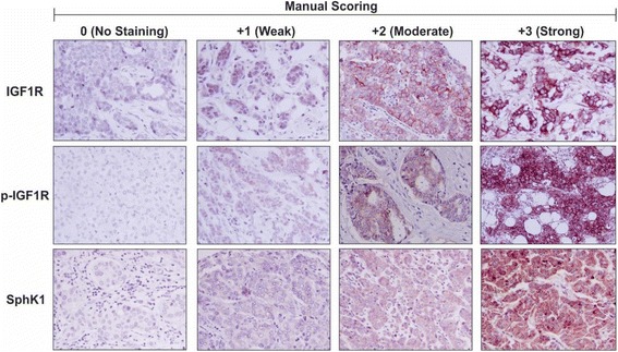
Fig. 1.

Immunohistochemistry and manual scoring analysis of Australian Breast Cancer Tissue Bank patient samples. Immunohistochemistry was performed on formalin-fixed paraffin embedded breast cancer patient tissue samples (n = 236) obtained from the Australian Breast Cancer Tissue Bank (ABCTB) using antibodies to detect and measure relative levels of IGF1R, p-IGF1R and SphK1. The intensity of immunostaining was assessed by manual scoring according to standard guidelines as follows: 0 = no staining, 1 = weak staining, 2 = moderate staining and 3 = strong staining for IGF1R, p-IGF1R and SphK1
