
Fig. 2.
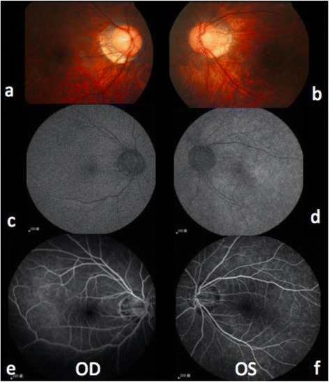
Binocular images of color fundoscopy, fundus autofluorescence, and fluorescein angiography on the next day after onset of symptoms. Color fundoscopy (a-b) revealed no specific finding except myopic crescent. Compared to the right eye (c), fundus autofluorescence revealed hyper-autofluorescence of the left eye (d). Fluorescein angiography (e-f) revealed retina small vessels vasculitis inflammation of the left eye(f)