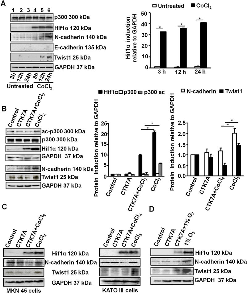
Fig. 1.
HAT inhibition downregulated CoCl2 and 1% O2-induced expression of metastatic markers in GCCs. (A) Expression of various metastatic factors was assessed at 3 h, 12 h, 24 h after treatment with 200 µM of CoCl2 by western blotting (n = 3). GAPDH was used as a loading control. Graphical presentation of western blot data clearly showed significant induction of Hif1α from 3 h to 24 h of CoCl2 treatment (mean ± SEM, n = 3), *P < 0.05. (B) Western blot analysis (n = 3) of whole cell lysates from AGS cells after treatment with 200 µM of CoCl2 and/or 100 µM of CTK7A for 24 h. Bar diagrams represent status of EMT markers (mean ± SEM). *P< 0.05 (C) Western blot analysis (n = 3) indicated downregulation of metastatic markers N-cadherin, Twist1, Hif1α in metastatic gastric cancer cell lines KATO III and MKN 45 after CoCl2 and CTK7A treatment. (D) Western blot analysis (n = 3) of whole cell lysates from AGS cells after a hypoxia (1% O2) exposure alone or with CTK7A. GAPDH was used as a loading control.

