Fig. 6.

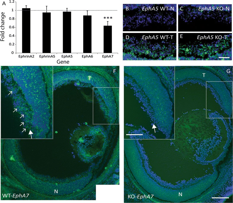
EphA7 is usually expressed in the ventrotemporal retinal crescent and is reduced in Ten-m3 KOs. A Realtime qPCR revealed significant down-regulation of EphA7 mRNA in the retina (fold change 0.64 ± 0.01, p < 0.001) of Ten-m3 KOs compared to WTs. No change in expression was detected in any of the other members of the EphA/ephrinA family tested. Graph shows relative fold change ± 1SE, normalised to Gapdh and in comparison to WT controls. Statistical significance is denoted by (*): p < 0.05*, p < 0.01**, p < 0.001***. B–E In situ hybridisation for EphA5 (green staining) on retinal sections for WT (B, D) and Ten-m3 KO (C, E) revealed no differences in expression pattern in the RGC layer. Signal was low in samples from nasal (N) retina from both WT and KO (B, C). Similarly high levels of expression observed in samples from temporal (T) retina of both genotypes (D, E). In situ hybridisation signal is superimposed on DAPI stain (blue) to reveal cell nuclei. Scale bar: 50 μm; applies to B–E. F, G In situ hybridisation on horizontal retinal sections for EphA7. In sections from WT (F), EphA7 (green staining) is expressed in a subset of cells in the RGC layer from far temporal (T) retina [dotted square; inset (large arrow)]. A fluorescent Nissl counterstain (DAPI) is shown in blue. Small arrows highlight a subset of EphA7 positive cells in this region. In an equivalent section from a Ten-m3 KO retina (G), EphA7 expression is markedly reduced, including in the RGC layer (arrow in inset). No EphA7 positive cells are visible in this region. Scale bar: 200 μm; scale in inset: 100 μm; applies to F and G. N nasal
