Figure 1.

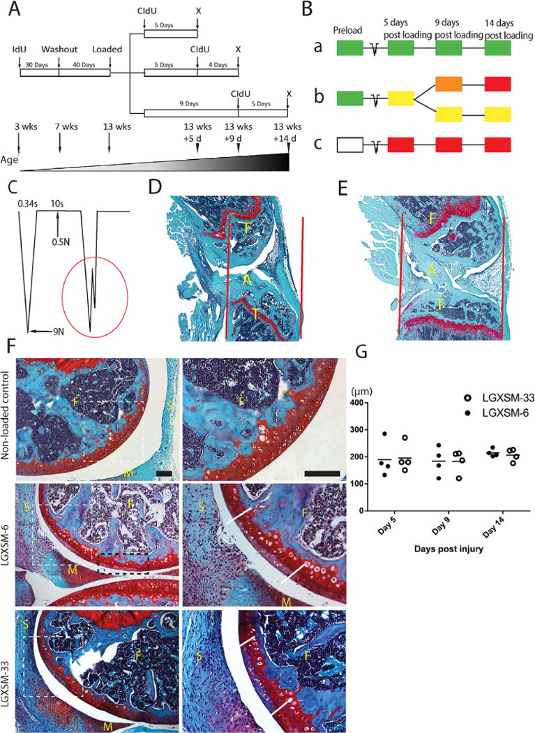
Non-invasive tibial compression and acute pathological changes in articular cartilage. (A) A sketch of double nucleoside cell-labeling procedure and (B) labeling options with three possible outcomes (a, b, and c). (a) IdU-positive cells will not proliferate and retain IdU (green); (b) IdU-positive cells will proliferate and acquire CIdU (red) and gradually lose IdU, hence appearing yellow, orange, or red; (c) Unlabeled IdU-negative cells (white) will proliferate and acquire CIdU (red); x = sacrifice. (C) A schematic diagram of loading cycle with basal maintenance force, peak force, time-interval between loading episodes and a typical indication of ACL rupture (red circle). (D) Compression-induced ACL rupture indicated by disrupted alignment of the ligament fibrils (A=ACL, F=femur, T=tibia), increased joint gap, and the dislocation of the tibia in relation to the femur; red lines indicate joint alignment. (E) Non-loaded control knee with intact ACL (A=ACL, F=femur, T=tibia). (F) Representative images of cartilage injury site of the lateral femoral condyle (left panel lower magnification, right panel higher magnification of white dotted line box), showing loss of Safranin-O staining and disappearance of normal nuclear staining. White dotted line box indicates the primary injury site, black dotted line box shows secondary injury site, parallel solid white lines indicate the injury border. F=femur, M=meniscus, S=synovium. Bar=100 μm. (G) Mean cartilage injury length showing no significant difference between the strains or time points. (n=4 each time point and each strain).
