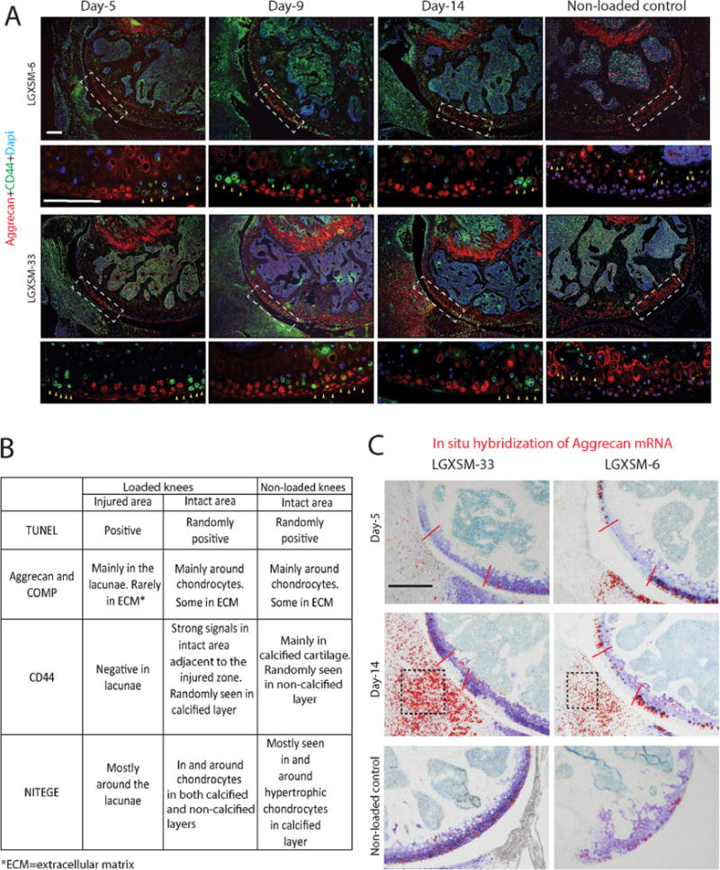
Figure 3.

CD44 immunofluorescence and in situ hybridization of aggrecan mRNA. (A) The areas indicated by white dotted line boxes in the upper panels are shown at higher magnification in the lower panels. CD44 (green) was seen around hypertrophic chondrocytes in non-loaded control knees (yellow arrows, the right panel) and in the chondrocytes immediately around the injured zone (yellow arrows, left three panels). There was no difference in the pattern of CD44 expression between the two mouse strains. Aggrecan staining (red) was undertaken to identify injury site. Bars=100 μm. (B) Summary of changes in extracellular matrix proteins in the injured and adjacent intact areas of loaded knees and in the intact area of non-loaded knees. (C) Aggrecan mRNA, detected by in situ hybridization, was only present in the intact area of cartilage and the cartilage of non-loaded knees at all time points in both strains (red color dots show positive hybridization signals, red lines show the injured area). Aggrecan mRNA was also seen in the synovium at day 14 post-injury in both strains (black boxes). Bar = 100 μm.
