
Figure 4. CD163 protein induction in THP-1 macrophages challenged with a single LPS stimulation.
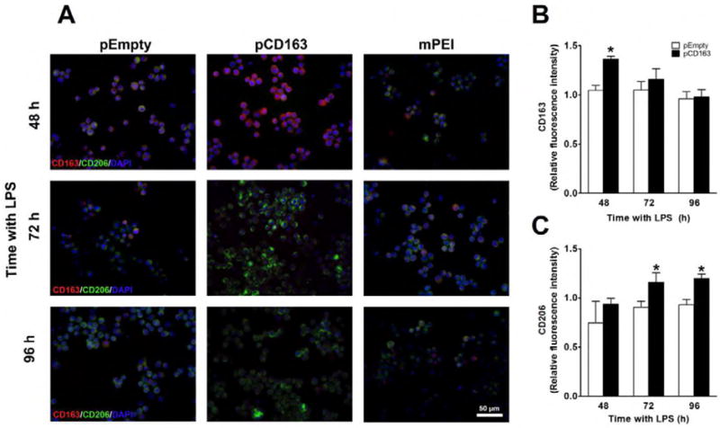
Microscopic images of nuclear staining using DAPI (blue), CD163 protein (red) and mannose receptor (CD206, green) in LPS-stimulated THP-1 macrophages transfected with Man-PEI complexed with an empty vector (pEmpty), a plasmid encoding for CD163 gene (pCD163) or the Man-PEI nanoparticle alone (A). Quantification of the relative average fluorescence intensity of CD163 (B) or mannose receptor (CD206, C) in LPS-stimulated THP-1 macrophages from 48 to 96 hours after transfection. The quantification of the relative average fluorescence intensity was normalized to the respective levels in the Man-PEI group, which was assigned a value equal to 1. N = 3 per group. *p< 0.05 vs. pEmpty by student’s t-test.