Fig. 1.

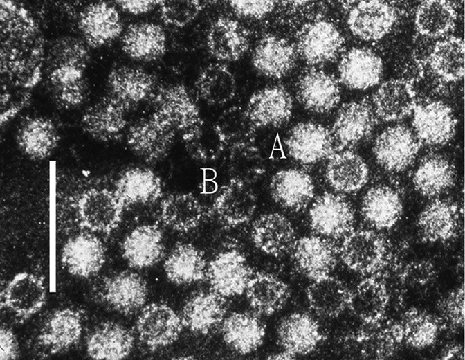
Negative contrast electron micrograph of human hepatitis E virus virions from a case stool collected in Nepal. (A) virion and (B) empty capsid. The bar represents 100 nm (photograph from M. Purdy).

Negative contrast electron micrograph of human hepatitis E virus virions from a case stool collected in Nepal. (A) virion and (B) empty capsid. The bar represents 100 nm (photograph from M. Purdy).