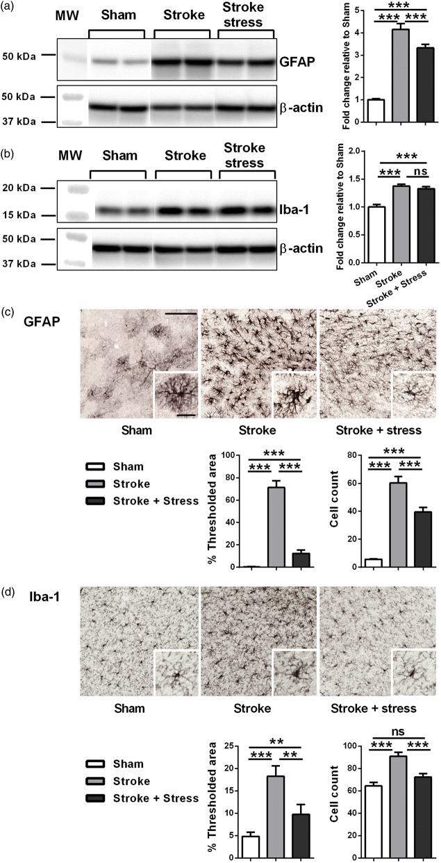
Figure 4.
Representative immunoblots for GFAP (a), Iba-1 (b), and β-actin from the peri-infarct region. The results for all the protein levels were calculated relative to β-actin levels. Data were expressed as a fold increase of the mean ± SEM for each group relative to the mean of the sham group. (n = 8 per group). ns: not significant, ***p < 0.016, Holm’s a priori analysis α correction procedure. Three panels on top illustrate representative labelling for GFAP (c) and Iba-1 (d) respectively for the three groups, sham, stroke and stroke + stress. Insets of c and d demonstrate the cellular structures of astrocyte and microglia, respectively. The bottom panels illustrate the quantification of the change in % of thresholded material and cell counts for each of the protein examined. Data expressed as mean ± SEM for sham = 8, stroke = 10 and stroke stress = 12. ns: not significant, ***p < 0.016, **p < 0.025, Holm’s a priori analysis α correction procedure. Bar on top of C represents 100 µm and bar of insets represents 30 µm.

