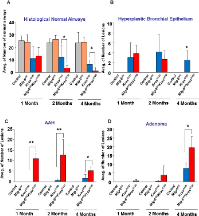
Figure 3. Mig-6 inactivation enhanced histopathological changes during KrasG12D–induced lung tumor progression.
Histopathological changes were scored in H&E stained sections of the lungs of control, Mig-6d/d, KrasG12D, and Mig-6d/dKrasG12D mice (at the age of 1, 2, and 4 months). Three mice were used for each group. Average numbers of normal airways (A), hyperplastic bronchial epithelium (B), Atypical Adenomatous Hyperplasia (AAH) (C), and adenomas (D) were calculated and shown in bar graphs. Independent pathologists counted and defined these histopathological patterns microscopically. *p<0.05 and **p<0.01 by One Way ANOVA followed by Tukey’s analysis.

