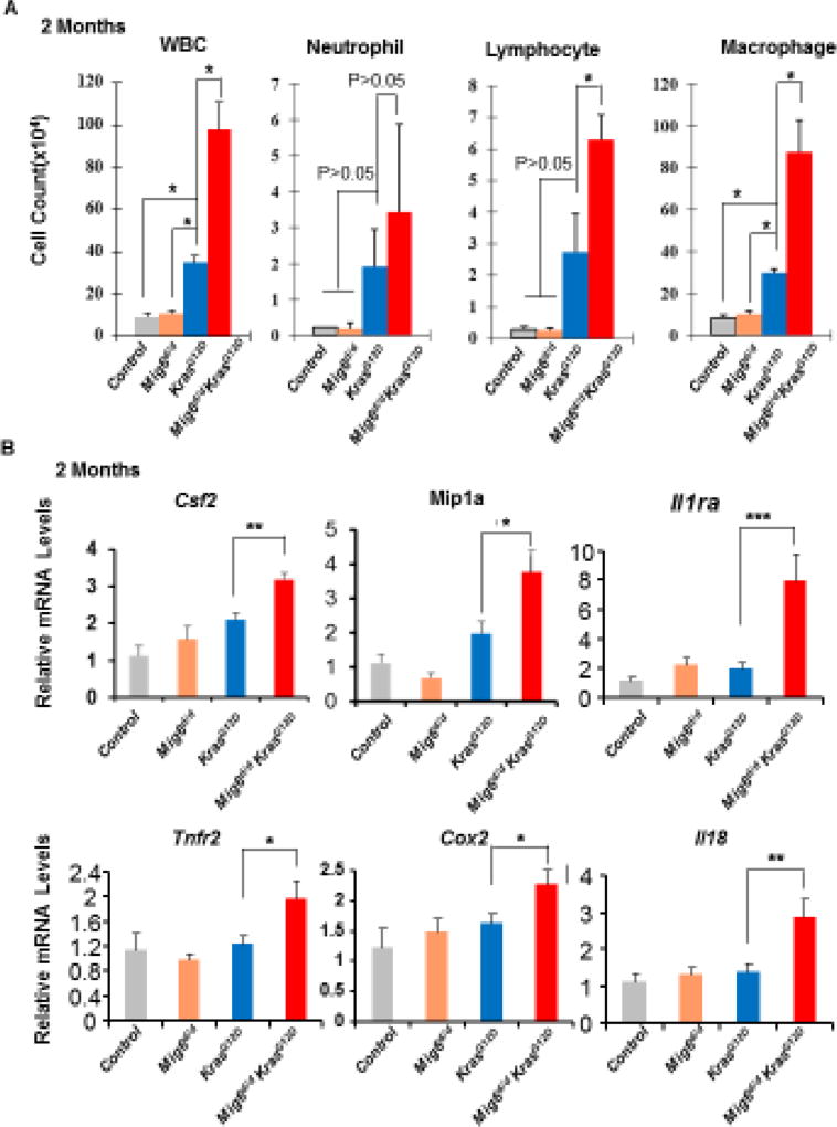
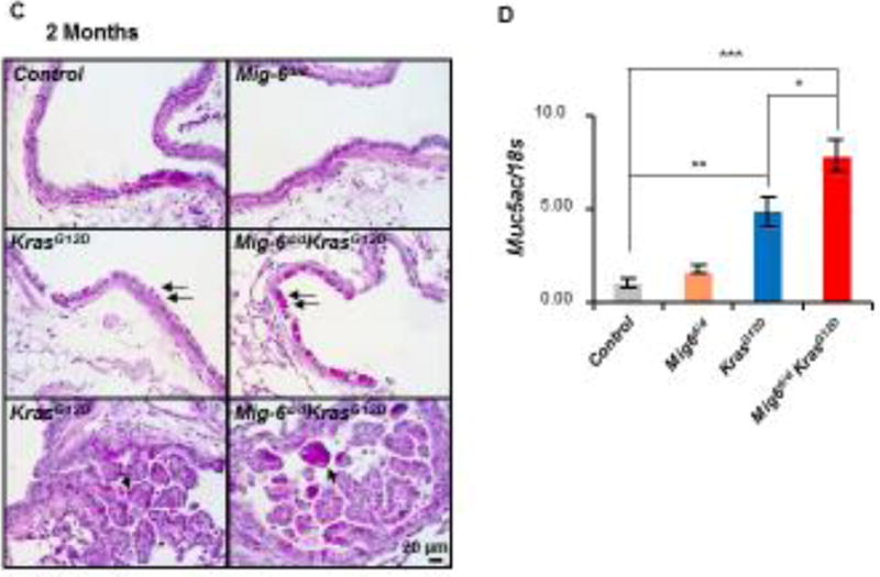
Figure 4. Inflammation was enhanced in Mig-6d/dKrasG12D mouse lungs.
(A) Analysis of white blood cells (WBC), neutrophils, lymphocytes, and macrophages in the BALF of control, Mig-6d/d, KrasG12D, and Mig-6d/dKrasG12D mice, *P<0.05. Three mice were used for each group. (B) RT-qPCR analysis of the expression of pro-inflammatory genes in the lungs of control, Mig-6d/d, KrasG12D, and Mig-6d/dKrasG12D mice. * p<0.05, ** p<0.01 and *** p<0.001. (C) PAS staining of lungs of control, Mig-6d/d, KrasG12D, and Mig-6d/dKrasG12D mice (2 months of ages). Arrows indicate metaplastic mucus cells in the bronchial epithelium. (D) RT-qPCR analysis of mRNA levels of Muc5ac in the lungs of mice (2 months of age). * p<0.05, ** p<0.01 and *** p<0.001.


