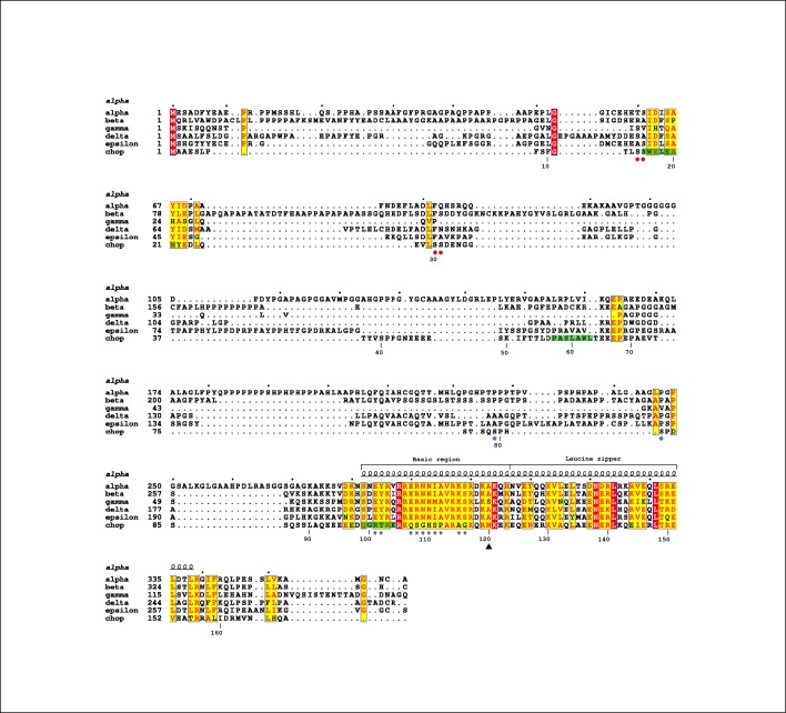
Fig 2. Alignment of the human C/EBP family.
Fully- and partially-conserved residues are in red and yellow, respectively. The secondary structure of the C/EBPα bZIP domain is shown above the sequences, while the numbering below corresponds to CHOP. The segments with α-helical propensity as identified by NMR (see below) are in green. Red and blue circles below the sequence indicate inactivating and activating phosphorylations in CHOP [7,8], while asterisks highlight unique changes in its DNA-binding domain. Figure prepared with ESPript (http://espript.ibcp.fr).

