Figure 2.

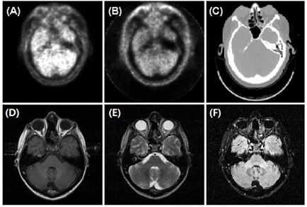
Two‐dimensional views of the common landmark (the skin) for PET with (a) and without (b) attenuation correction, CT (c) and MRI: T1 (d) T2 (e) and FLAIR (f).

Two‐dimensional views of the common landmark (the skin) for PET with (a) and without (b) attenuation correction, CT (c) and MRI: T1 (d) T2 (e) and FLAIR (f).