
Figure 4.
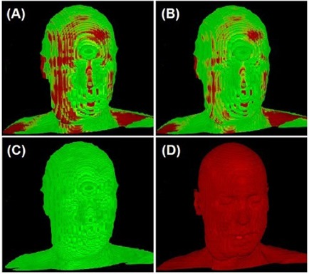
Correction of misalignment in a co‐registered PET/CT image: before (a) and after (b) the 3DVIR correction. The volumetric skin landmark in PET and CT images are shown in (c) and (d), respectively. In PET, the normal tissue uptake of can indicate some specific anatomical structures, which were used for registration, as shown in (c). Note that the best volumetric match would show most homogeneity of the color distribution on the skin landmark.